
APMAP
APMAP全稱為脂肪細胞質膜相關蛋白,是定位于脂肪細胞膜上的蛋白質,目前沒有特別廣泛使用的別名。它和脂肪組織的正常功能密切相關,比如參與脂肪細胞的分化、脂質的轉運等過程,幫助維持身體代謝的平衡。APMAP表達或功能出現異常時,可能會影響脂肪代謝的穩定,進而與肥胖、2型糖尿病等代謝性疾病產生關聯——臨床觀察發現,部分患者體內該蛋白的水平與疾病進展存在一定聯系。在藥物研發方面,針對它的研究仍處于早期階段,科研人員正探索其作為代謝疾病治療靶點的可能性,不過目前還沒有相關藥物進入臨床試驗階段。
熱銷產品
Recombinant Human Adipocyte plasma membrane-associated protein (APMAP), partial (CSB-EP884629HUb1)
驗證數據
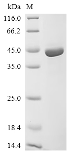
(Tris-Glycine gel) Discontinuous SDS-PAGE (reduced) with 5% enrichment gel and 15% separation gel.
APMAP Monoclonal Antibody (CSB-MA943823)
驗證數據
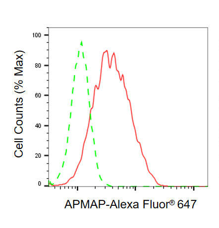
Flow cytometric analysis of APMAP expression in HT-1080 cells using APMAP antibody. Green, isotype control; red, APMAP.
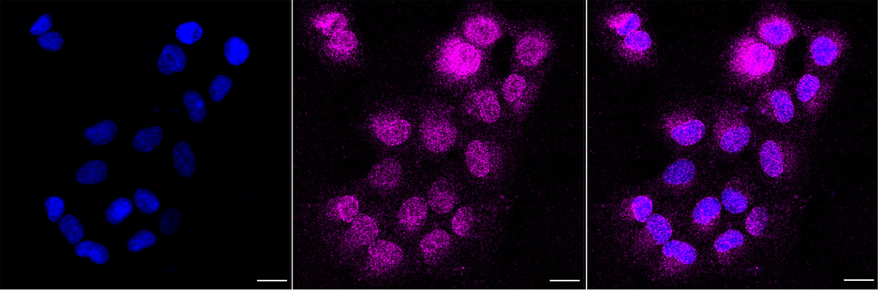
Immunocytochemical staining of HT-1080 cells with APMAP antibody. Nuclei were stained blue with DAPI; APMAP was stained magenta with Alexa Fluor? 647. Images were taken using Leica stellaris 5. Protein abundance based on laser Intensity and smart gain: Medium. Scale bar, 20 μm.
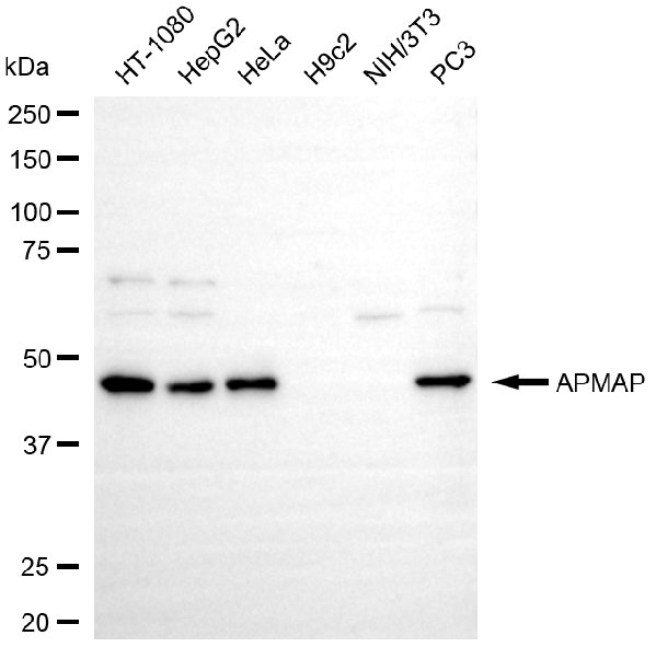
Western blotting analysis using APMAP antibody. Total cell lysates (30 μg) from various cell lines were loaded and separated by SDS-PAGE. The blot was incubated with APMAP antibody and HRP-conjugated goat anti-mouse secondary antibody respectively.
APMAP Antibody (CSB-PA884629LA01HU)
驗證數據
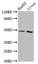
Western Blot
Positive WB detected in: HepG2 whole cell lysate, Rat liver tissue
All lanes: APMAP antibody at 3.1µg/ml
Secondary
Goat polyclonal to rabbit IgG at 1/50000 dilution
Predicted band size: 47, 33 kDa
Observed band size: 47 kDa
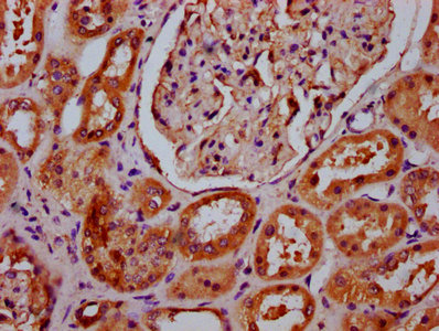
IHC image of CSB-PA884629LA01HU diluted at 1:400 and staining in paraffin-embedded human kidney tissue performed on a Leica BondTM system. After dewaxing and hydration, antigen retrieval was mediated by high pressure in a citrate buffer (pH 6.0). Section was blocked with 10% normal goat serum 30min at RT. Then primary antibody (1% BSA) was incubated at 4°C overnight. The primary is detected by a biotinylated secondary antibody and visualized using an HRP conjugated SP system.
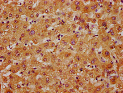
IHC image of CSB-PA884629LA01HU diluted at 1:400 and staining in paraffin-embedded human liver tissue performed on a Leica BondTM system. After dewaxing and hydration, antigen retrieval was mediated by high pressure in a citrate buffer (pH 6.0). Section was blocked with 10% normal goat serum 30min at RT. Then primary antibody (1% BSA) was incubated at 4°C overnight. The primary is detected by a biotinylated secondary antibody and visualized using an HRP conjugated SP system.
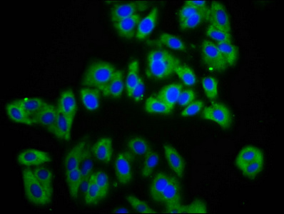
Immunofluorescence staining of HepG2 cells with CSB-PA884629LA01HU at 1:133, counter-stained with DAPI. The cells were fixed in 4% formaldehyde, permeabilized using 0.2% Triton X-100 and blocked in 10% normal Goat Serum. The cells were then incubated with the antibody overnight at 4°C. The secondary antibody was Alexa Fluor 488-congugated AffiniPure Goat Anti-Rabbit IgG(H+L).
APMAP Antibodies
APMAP for Homo sapiens (Human)
| 產品貨號 | 產品名稱 | 種屬反應性 | 應用類型 |
|---|---|---|---|
| CSB-PA165039 | APMAP Antibody | Human | ELISA,WB,IHC |
| CSB-PA915948 | APMAP Antibody | Human | ELISA,WB,IHC |
| CSB-PA884629LA01HU | APMAP Antibody | Human, Rat | ELISA, WB, IHC, IF |
| CSB-MA943823 | APMAP Monoclonal Antibody | Human | ELISA, WB, FC, ICC |
APMAP Proteins
APMAP Proteins for Homo sapiens (Human)
| 產品貨號 | 產品名稱 | 來源 |
|---|---|---|
| CSB-YP884629HU CSB-EP884629HU CSB-BP884629HU CSB-EP884629HU-B |
Recombinant Human Adipocyte plasma membrane-associated protein (APMAP), partial | Yeast E.coli Baculovirus In Vivo Biotinylation in E.coli |
| CSB-EP884629HUb1 | Recombinant Human Adipocyte plasma membrane-associated protein (APMAP), partial | E.coli |
| CSB-MP884629HU | Recombinant Human Adipocyte plasma membrane-associated protein (APMAP), partial | Mammalian cell |
| CSB-MP884629HUd7 | Recombinant Human Adipocyte plasma membrane-associated protein (APMAP), partial | Mammalian cell |