DLAT
DLAT,全稱為二氫硫辛酸轉乙酰基酶,常被稱為丙酮酸脫氫酶復合物E2亞基(PDC-E2)。它是細胞能量代謝里的關鍵分子,能幫助把糖分解產生的丙酮酸轉化為乙酰輔酶A,為身體獲取能量提供重要支持。
在疾病方面,DLAT和原發性膽管炎關系密切,多數患者體內存在針對PDC-E2的自身抗體,這些抗體可能影響它的正常工作,引發膽管炎癥。此外,DLAT功能異常還可能導致丙酮酸脫氫酶缺乏癥,患者常出現肌肉無力、發育遲緩等問題。目前針對相關疾病的治療,原發性膽管炎常用熊去氧膽酸緩解癥狀;針對代謝缺陷的輔助治療包括補充輔酶類物質,但專門靶向DLAT的藥物仍在早期研發階段,比如調節其活性的小分子化合物,尚未進入臨床應用。
熱銷產品
驗證數據
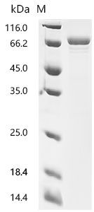
(Tris-Glycine gel) Discontinuous SDS-PAGE (reduced) with 5% enrichment gel and 15% separation gel.
DLAT Recombinant Monoclonal Antibody (CSB-RA283972A0HU)
驗證數據
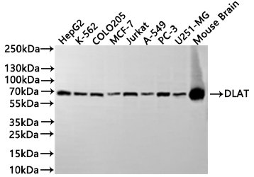
Western Blot
Positive WB detected in: HepG2 whole cell lysate(30μg), K562 whole cell lysate(30μg), COLO205 whole cell lysate(30μg), MCF7 whole cell lysate(30μg), Jurkat whole cell lysate(30μg), A549 whole cell lysate(30μg), PC-3 whole cell lysate(30μg), U251 whole cell lysate(30μg), Mouse brain tissue lysate(30μg)
All lanes:DLAT antibody at 1:1000
Secondary
Goat polyclonal to rabbit IgG at 1/40000 dilution
Predicted band size: 69 kDa
Observed band size: 69 kDa
Exposure time:2min
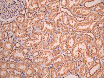
IHC image of CSB-RA283972A0HU diluted at 1:100 and staining in paraffin-embedded human kidney tissue performed on a Leica BondTM system. After dewaxing and hydration, antigen retrieval was mediated by high pressure in a citrate buffer (pH 6.0). Section was blocked with 10% normal goat serum 30min at RT. Then primary antibody (1% BSA) was incubated at 4°C overnight. The primary is detected by a Goat anti-rabbit polymer IgG labeled by HRP and visualized using 0.05% DAB.
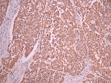
IHC image of CSB-RA283972A0HU diluted at 1:100 and staining in paraffin-embedded human liver cancer performed on a Leica BondTM system. After dewaxing and hydration, antigen retrieval was mediated by high pressure in a citrate buffer (pH 6.0). Section was blocked with 10% normal goat serum 30min at RT. Then primary antibody (1% BSA) was incubated at 4°C overnight. The primary is detected by a Goat anti-rabbit polymer IgG labeled by HRP and visualized using 0.05% DAB.
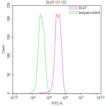
Overlay Peak curve showing MCF-7 cells stained with CSB-RA283972A0HU (red line) at 1:100. The cells were fixed in 4% formaldehyde and permeated by 0.2% TritonX-100 for 10min. Then 10% normal goat serum to block non-specific protein-protein interactions followed by the antibody (1ug/1*106cells) for 45min at 4℃. The secondary antibody used was FITC-conjugated goat anti-rabbit IgG (H+L) at 1/200 dilution for 35min at 4℃.Control antibody (green line) was Rabbit IgG (1ug/1*106cells) used under the same conditions. Acquisition of >10, 000 events was performed.
DLAT Antibody (CSB-PA006926LA01HU)
驗證數據
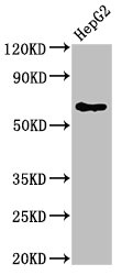
Western Blot
Positive WB detected in: HepG2 whole cell lysate
All lanes: DLAT antibody at 4μg/ml
Secondary
Goat polyclonal to rabbit IgG at 1/50000 dilution
Predicted band size: 69 kDa
Observed band size: 69 kDa
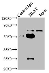
Immunoprecipitating DLAT in HepG2 whole cell lysate
Lane 1: Rabbit control IgG instead of CSB-PA006926LA01HU in HepG2 whole cell lysate.
For western blotting, a HRP-conjugated Protein G antibody was used as the secondary antibody (1/2000)
Lane 2: CSB-PA006926LA01HU (6μg) + HepG2 whole cell lysate (1mg)
Lane 3: HepG2 whole cell lysate (20μg)
DLAT Antibodies
DLAT for Homo sapiens (Human)
| 產品貨號 | 產品名稱 | 種屬反應性 | 應用類型 |
|---|---|---|---|
| CSB-PA006926GA01HU | DLAT Antibody | Human,Mouse,Rat | ELISA,WB |
| CSB-PA445587 | DLAT Antibody | Human,Mouse,Rat | ELISA,WB,IHC |
| CSB-PA084734 | DLAT Antibody | Human,Mouse,Rat | ELISA,WB,IHC |
| CSB-PA006926LA01HU | DLAT Antibody | Human | ELISA, WB, IP |
| CSB-RA283972A0HU | DLAT Recombinant Monoclonal Antibody | Human, Mouse | ELISA, WB, IHC, FC |
| CSB-RA238917A0HU | DLAT Recombinant Monoclonal Antibody | Human, Mouse, Rat | ELISA, WB, FC, ICC, IHC |
| CSB-RA441447A0HU | DLAT Recombinant Monoclonal Antibody | Human, Mouse, Rat | ELISA, WB, FC, ICC |
DLAT Proteins
DLAT Proteins for Homo sapiens (Human)
| 產品貨號 | 產品名稱 | 來源 |
|---|---|---|
| CSB-YP006926HU CSB-BP006926HU CSB-MP006926HU CSB-EP006926HU-B |
Recombinant Human Dihydrolipoyllysine-residue acetyltransferase component of pyruvate dehydrogenase complex, mitochondrial (DLAT) | Yeast Baculovirus Mammalian cell In Vivo Biotinylation in E.coli |
| CSB-EP006926HU | Recombinant Human Dihydrolipoyllysine-residue acetyltransferase component of pyruvate dehydrogenase complex, mitochondrial (DLAT), partial | E.coli |
| CSB-EP006926HUc7 | Recombinant Human Dihydrolipoyllysine-residue acetyltransferase component of pyruvate dehydrogenase complex, mitochondrial (DLAT), partial | E.coli |
DLAT Proteins for Mus musculus (Mouse)
| 產品貨號 | 產品名稱 | 來源 |
|---|---|---|
| CSB-YP804374MO | Recombinant Mouse Dihydrolipoyllysine-residue acetyltransferase component of pyruvate dehydrogenase complex, mitochondrial (Dlat) | Yeast |
| CSB-EP804374MO CSB-BP804374MO CSB-EP804374MO-B |
Recombinant Mouse Dihydrolipoyllysine-residue acetyltransferase component of pyruvate dehydrogenase complex, mitochondrial (Dlat) | E.coli Baculovirus In Vivo Biotinylation in E.coli |
| CSB-MP804374MO | Recombinant Mouse Dihydrolipoyllysine-residue acetyltransferase component of pyruvate dehydrogenase complex, mitochondrial (Dlat) | Mammalian cell |
| CSB-EP804374MOa0 | Recombinant Mouse Dihydrolipoyllysine-residue acetyltransferase component of pyruvate dehydrogenase complex, mitochondrial (Dlat) | E.coli |
| CSB-MP804374MOe1 | Recombinant Mouse Dihydrolipoyllysine-residue acetyltransferase component of pyruvate dehydrogenase complex, mitochondrial (Dlat) | Mammalian cell |